Understanding Cell Culture Plate Geometry in Assay Design
Selecting the correct microplate geometry is a critical yet frequently overlooked parameter in cell-based assay optimization and general assay sensitivity optimization. Microplate well architecture dictates fluid dynamics, light transmission, and biological spatial distribution within every assay well. Understanding cell culture plate geometry variables is essential for minimizing background noise, improving cell attachment or cell aggregation dynamics, and maximizing overall experimental precision.
The Role of Plate Geometry in Assay Precision
Well bottom shape alters light reflection patterns and modifies how adherent or suspension cells settle across the culture surface.
- Light Path Uniformity: Flat surfaces deliver a constant, parallel optical path length across the entire bottom area, preventing lens-like light refraction.
- Cell Distribution: Flat planes encourage uniform monolayer coverage and stable cell attachment, whereas curved profiles force cells toward the central vortex to promote intentional cell aggregation.
- Signal-to-Noise Ratio (SNR): Proper cell culture plate selection minimizes light scatter and edge effects, boosting signal clarity during measurement.
Introducing Puretest Laboratory Solutions
Puretest delivers high-grade consumable solutions manufactured under strict quality standards to empower precise diagnostic and therapeutic research.
- Class 100K Cleanroom Production: All flat-bottom cell culture plates and U-bottom cell culture plates are molded in ISO-compliant facilities to guarantee pyrogen-, DNase-, and RNase-free standards.
- Advanced Vacuum-Gas Plasma TC-Treatment: Surface modification across every TC-treated cell culture plate ensures highly consistent hydrophilicity for reproducible cell attachment.
- Custom OEM/ODM Engineering: Tailored molding and packaging configurations designed for automated screening platforms.
Comparative Analysis: Flat Bottom vs U-Bottom Cell Culture Plates
Choosing between flat bottom vs U-bottom cell culture plates depends fundamentally on whether your protocol relies on monolayer observation or cell concentration.
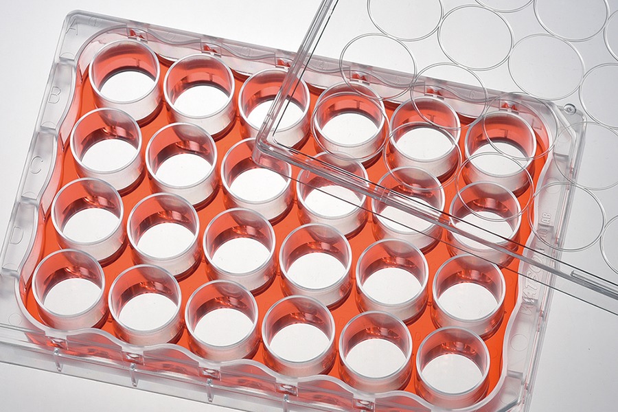
Flat Bottom Cell Culture Plates: Maximizing Surface Area and Optical Uniformity
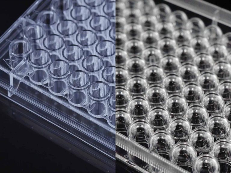
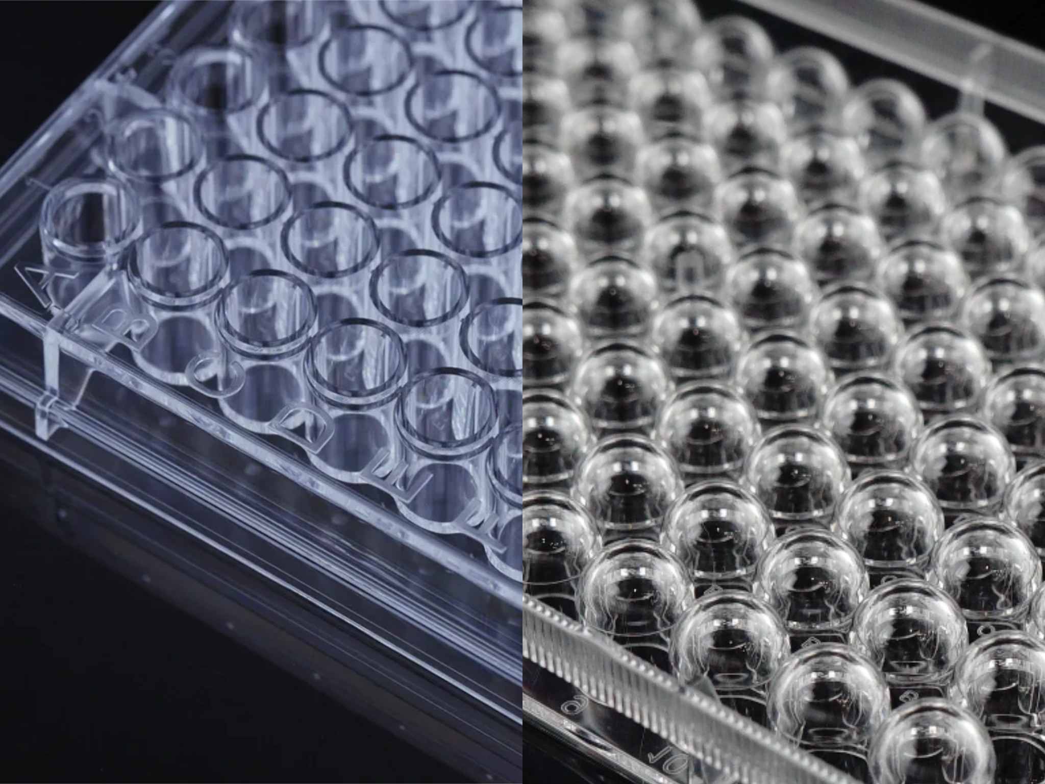
Flat bottom cell culture plates offer a planar surface that ensures uniform light path lengths across the entire well bottom.
- High Optical Clarity: Premium virgin polystyrene construction delivers superior optical clarity while minimizing light scattering during spectrophotometric reading.
- Optimal Anchorage Surface: Provides maximum growth surface area (approx. 0.32 cm² in 96-well formats) for reliable cell attachment of adherent cell lines like HeLa, HEK293, and NIH/3T3.
- Microscopy Compatibility: Utilizing a flat bottom plate for microscopy eliminates focal field distortions, permitting clear high-content screening (HCS) from well center to margin.
U-Bottom Cell Culture Plates: Enforcing Cell Aggregation and Liquid Recovery
U-bottom cell culture plates (round bottom) feature a hemispherical base that directs gravitationally settling particles to a single focal point.
- Spheroid Aggregation: Guides non-adherent cells or specialized stem cells to coalesce into 3D embryoid bodies through efficient cell aggregation.
- Concentrated Pelleting: Ideal for washing suspension cultures or executing centrifugation-based immunoassays.
- Low Dead Volume: Allows complete liquid evacuation during automated washing procedures, preserving valuable reagents.
Impact of Well Geometry on Specific Assay Readouts
Detection hardware works together with well bottom geometry. This shapes cell-based assay optimization, along with the standard deviations that show up in experiments and the CV numbers that follow.
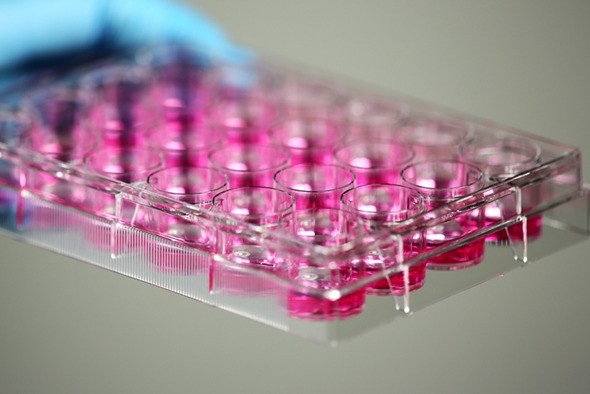
Spectrophotometric and Microscopic Assays (Flat Bottom Dominance)
Plate readers and automated microscopes depend on steady light paths. They produce solid absorbance and fluorescence numbers as a result.
- Absorbance Assays (MTT/CCK-8): Flat-bottom cell culture plates ensure consistent liquid depth, preventing light beam distortion and maintaining Beer-Lambert law compliance (improving assay CV by up to 15%).
- Fluorescence and Luminescence: The fluorophores spread evenly through the liquid. This cuts down on local quenching and stops central signal spikes from forming.
- Automated Imaging: Relying on a flat bottom plate for microscopy keeps focal planes steady across the full view. It shortens autofocus time when high-throughput scanners run their scans.
3D Spheroid and Immunoassay Workflows (U-Bottom Advantages)
Curved geometries in U-bottom cell culture plates are tailored for protocols requiring localized particle accumulation or cell-to-cell interaction.
- Tumor Spheroid Models: Round bottoms facilitate consistent single-spheroid assembly per well via controlled cell aggregation, essential for 3D drug toxicity profiling.
- Agglutination & Bead Assays: Concentrates magnetic beads or red blood cells into a tight central spot for rapid visual or digital scoring.
- Sample Extraction Efficiency: Minimizes residual volume during washing steps, boosting signal purity in ELISAs.
Puretest High-Performance Flat Bottom Cell Culture Plates
To ensure maximal optical transmittances and cellular adhesion, Puretest manufactures a versatile range of flat bottom cell culture plates designed for rigorous scientific applications.
Puretest 96-Well TC-Treated Flat Bottom Culture Plate
Engineered specifically for high-throughput screening, enzyme-linked assays, and quantitative colorimetric evaluations.
- Flatness Tolerances: Bottom planarity deviation maintained within strictly controlled tolerances to comply with ANSI/SLAS 1-4 standards.
- Advanced TC-Surface: As a top-tier TC-treated cell culture plate, uniform negative charge density enhances cell attachment rates, yielding monolayer uniformity across all 96 wells.
- Application Focus: Resolves uneven signal detection in MTT and CCK-8 proliferation assays by delivering flat light path lengths and low inter-well CV (< 3.5%).
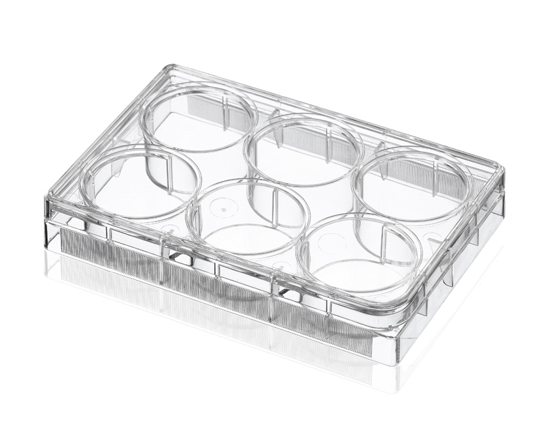
Puretest 24-Well and 6-Well TC-Treated Flat Bottom Plates
Designed for expanded primary cell cultures, live-cell imaging, and transwell-style functional assays.
- High Contrast Clarity: Ultra-clear medical-grade polystyrene ensures exceptional optical clarity during phase-contrast and fluorescent microscopy.
- Serrated Rim & Lidded Venting: Molded anti-evaporation rings and raised well rims eliminate cross-contamination and edge-effect liquid loss.
- Application Focus: Using a TC-treated cell culture plate with flat-bottom geometry solves image blurring during high-magnification microscopy, allowing precise focal stacking in live-cell migration and transfection assays.
Strategic Guidelines for Selecting the Optimal Plate Geometry
Aligning protocol requirements with cell culture plate geometry prevents costly experimental re-runs and technical artifacts.
Decision Matrix for Assay Sensitivity Optimization
Refer to this quick cell culture plate selection workflow when planning your experimental protocols:
- Select Flat Bottom Cell Culture Plates When: Conducting absorbance-based cell viability, direct ELISA, fluorescent microplate reading, or automated optical imaging on adherent monolayers requiring strong cell attachment.
- Select U-Bottom Cell Culture Plates When: Culturing 3D tumor spheroids through cell aggregation, performing cell suspension centrifugation, or running agglutination assays requiring minimal dead volumes.
- Assess Surface Energy Needs: Ensure a TC-treated cell culture plate is selected for adherent lines, while non-treated options are reserved for suspension or spheroid protocols.
Conclusion
Achieving successful cell-based assay optimization and overall assay sensitivity optimization requires balancing biological culture needs with instrument optical requirements. In the evaluation of flat-bottom vs U-bottom cell culture plates, while U-bottom plates excel at 3D cell aggregation and suspension recovery, Puretest flat-bottom cell culture plates deliver the optical clarity, planar surface uniformity, and consistent cell attachment required for rigorous quantitative assays.
Ready to eliminate optical artifacts and elevate your lab’s assay precision? Make the right cell culture plate selection today—contact Puretest to request free sample kits or discuss custom OEM/ODM microplate manufacturing tailored to your exact screening specs!
FAQ
Q: How does cell culture plate geometry affect absorbance readings in colorimetric cell viability assays?
A: Flat bottom cell culture plates ensure a uniform liquid path length across the light beam. U-bottom cell culture plates create a concave liquid meniscus that refracts light unpredictably, causing significant optical variance and elevated CV values.
Q: Can U-bottom cell culture plates be used for adherent cell culture?
A: While adherent cells can attach to a TC-treated cell culture plate with a U-bottom surface, gravity forces them to accumulate densely at the center. This causes local over-confluence, nutrient depletion, and severe distortion when a flat-bottom plate for microscopy is required instead.
Q: Why are flat bottom cell culture plates preferred for automated high-content screening (HCS)?
A: A flat bottom plate for microscopy maintains a uniform focal plane across the entire surface area. This allows automated microscope optics to lock focus rapidly without constant z-axis recalibration between well centers and edges.
Q: How does Puretest TC-treatment enhance cell monolayer consistency in flat bottom cell culture plates?
A: Every Puretest TC-treated cell culture plate utilizes controlled vacuum-gas plasma treatment to incorporate oxygen-containing functional groups onto the polystyrene surface. This increases surface hydrophilicity, promoting uniform cell attachment and preventing cell clumping.